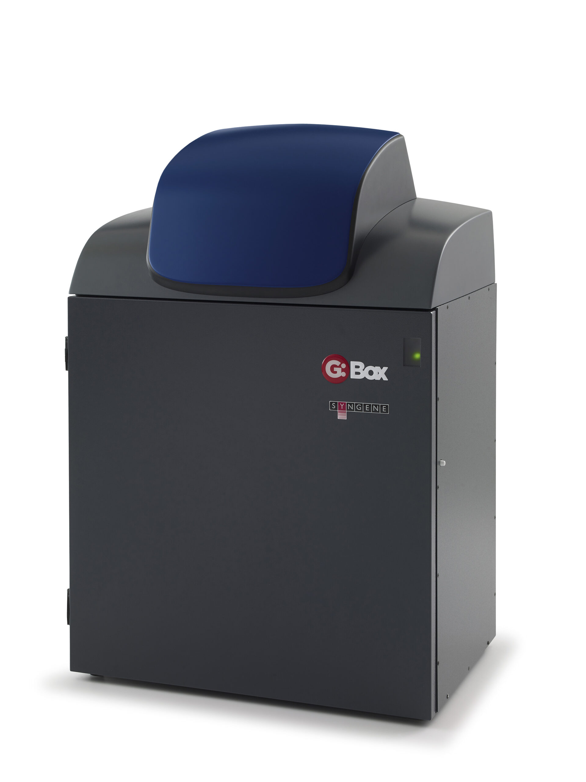
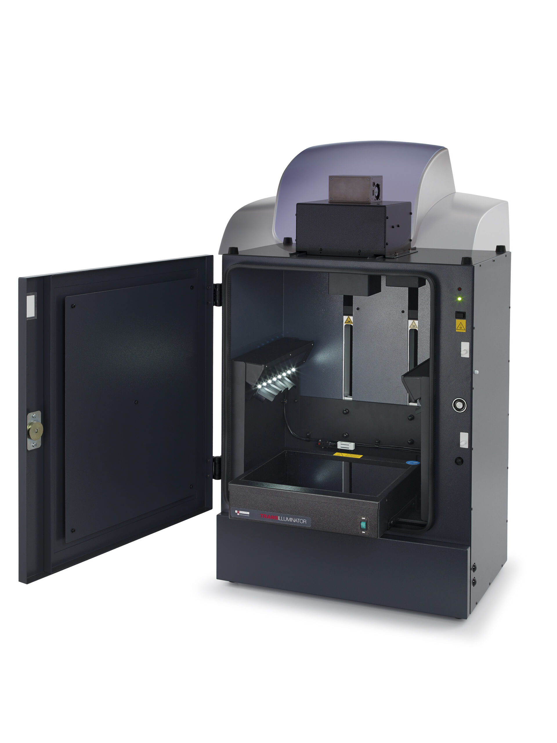
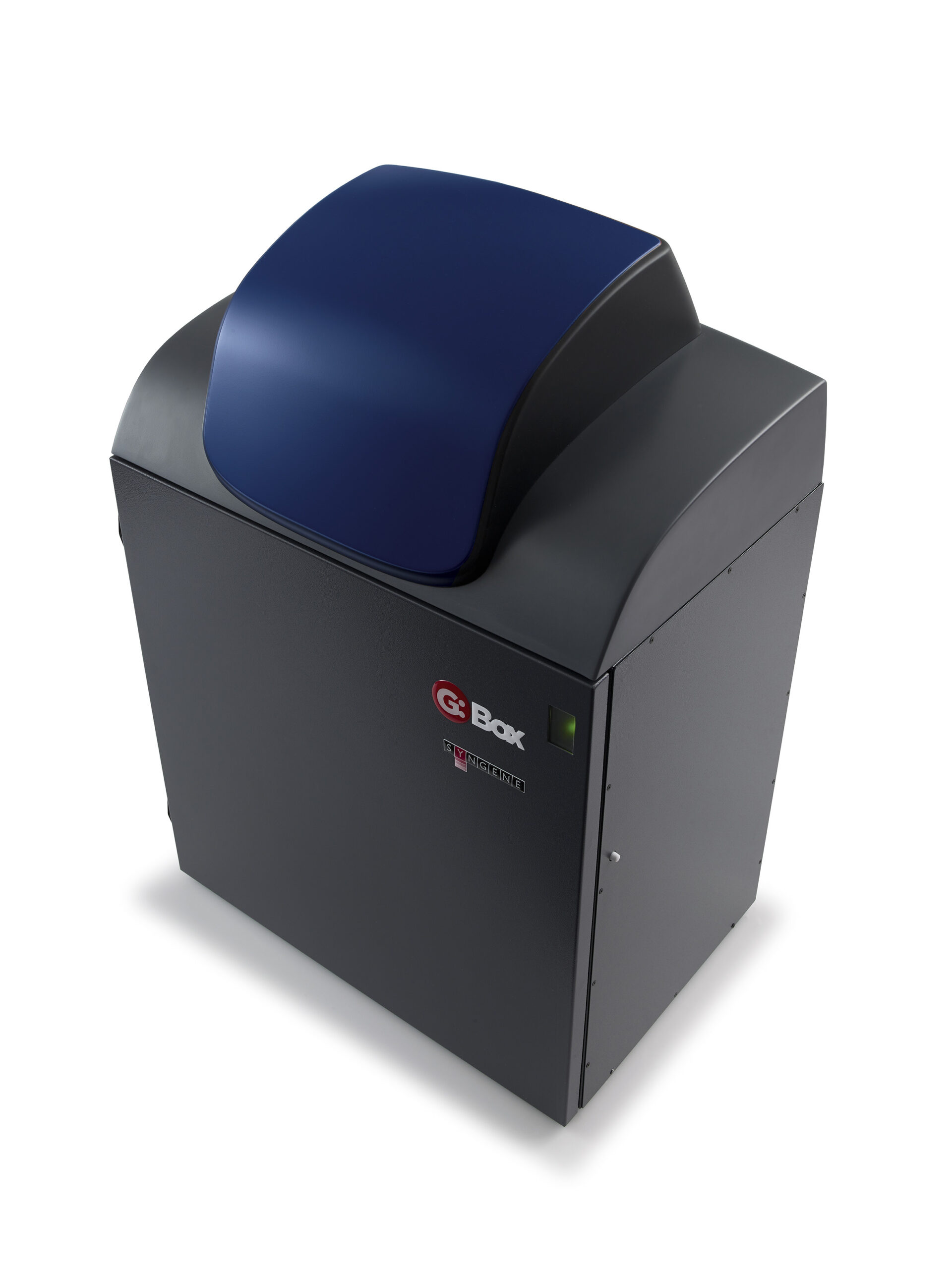
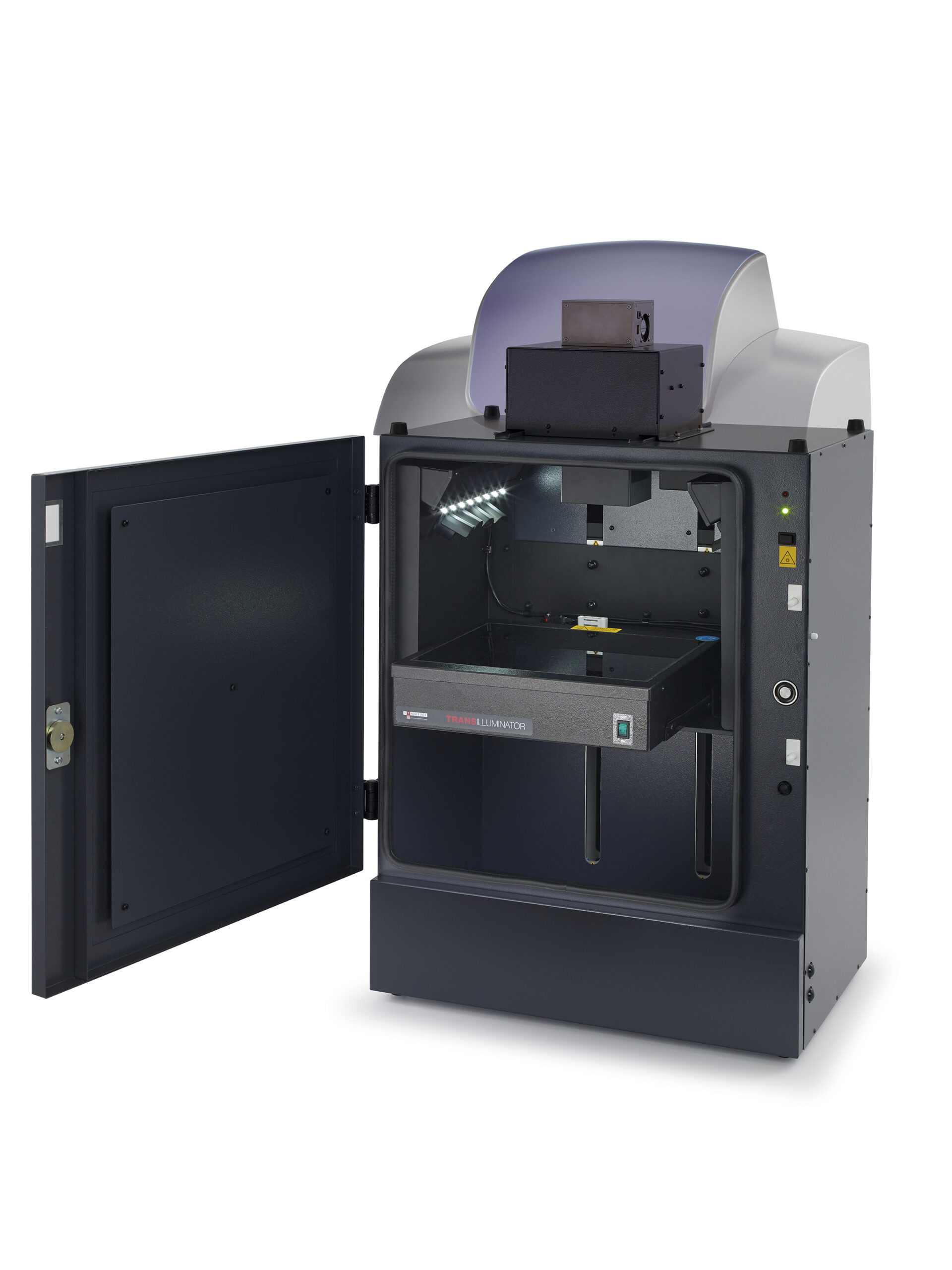
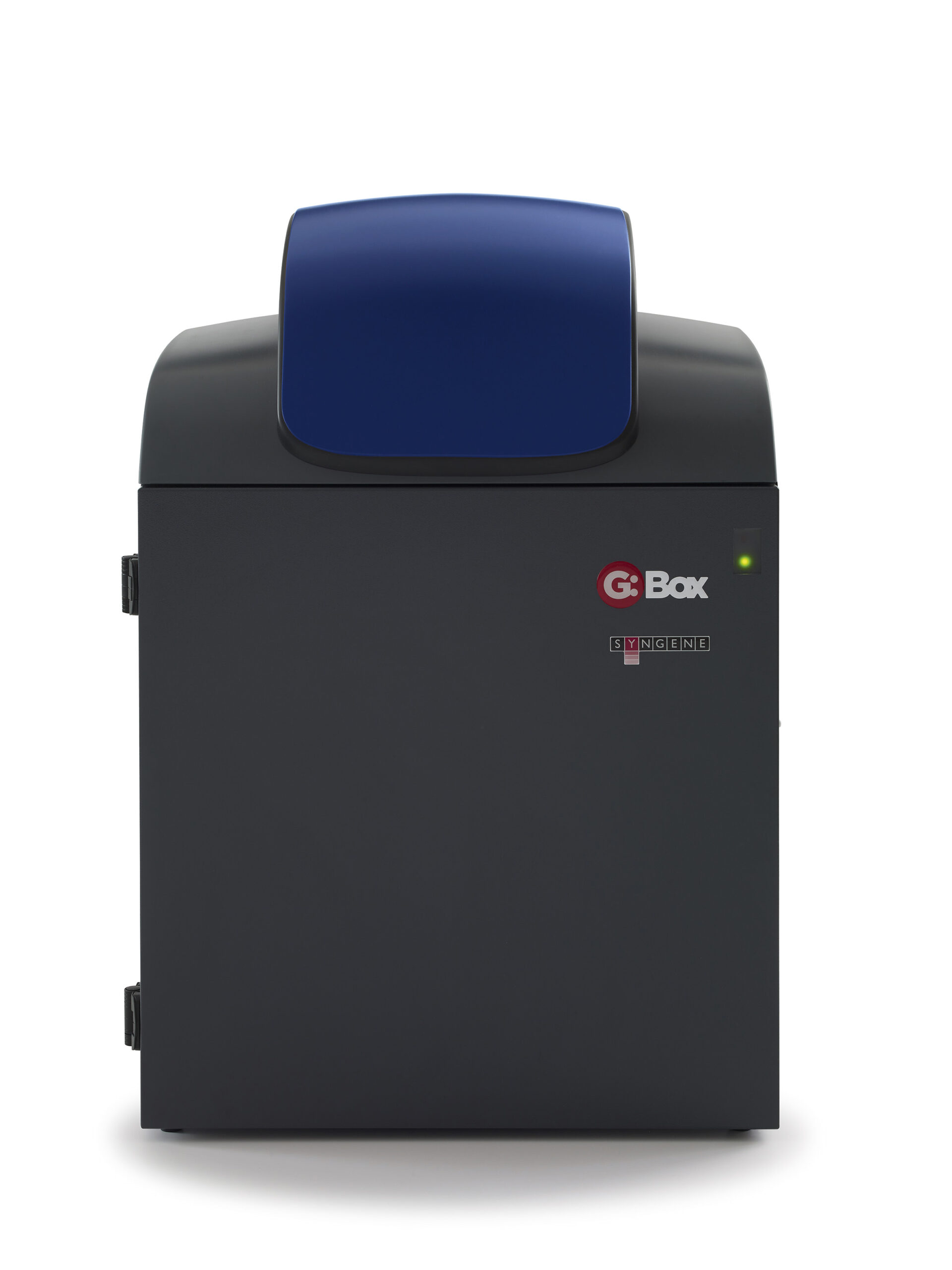
Your lab’s imaging system shouldn’t control how you detect proteins on Western blots. Chemiluminescence is great if you want sensitive detection of picogram or femtogram amounts, while fluorescence lets you quantify and detect multiple different proteins on one blot. With a G:BOX Chemi XX6/XX9 you can have it all. Powered by Syngene’s revolutionary GeneSys software featuring hundreds of imaging protocols, you’re free to choose chemiluminescence and fluorescence reagents from any manufacturer, putting you in charge of how you detect your proteins.
Featuring an extended darkroom, you can choose between a 6 or 9 mega pixel, cooled, high quantum efficiency camera for unrivaled levels of sensitivity with minimal background interference.
HI-LED lighting options cover the full spectrum of high intensity blue, green, red and infra-red resulting in faster exposure times and publication quality images. An edge lighting option can also be used for 2D gel capture including the use of DIGE gels.
The system is controlled by GeneSys application driven image capture software and comes complete with unlimited copies of GeneTools analysis software.
| G:BOX Chemi XX6 | G:BOX Chemi XX9 | |
|---|---|---|
| Image resolution (pixels m) | 6 | 9.1 |
| Effective resolution (pixels m) | 18 | 27 |
| A/D | 16 bit | 16 bit |
| Greyscales | 65536 | 65536 |
| Quantum efficiency @ 425nm | 73% | 73% |
| Cooling | Peltier | Peltier |
| Lens (motor driven) | Motor driven f0.95 with auto focus and stage zoom |
Motor driven f0.95 with auto focus and stage zoom |
| Filter wheel (7 position motor driven) | Yes | Yes |
| UV filter | Yes | Yes |
| Use with external PC | Yes | Yes |
| Darkroom | ||
| Extended with motor driven stage | Yes | Yes |
| Illumination | ||
| Epi LED white lights | Yes | Yes |
| Epi UV 302nm | Optional | Optional |
| Epi red LED module | Optional | Optional |
| Epi blue LED module | Optional | Optional |
| Epi green LED module | Optional | Optional |
| Epi red LED module M series for multiplexing | Optional | Optional |
| Epi green LED module M series for multiplexing | Optional | Optional |
| Epi blue LED module M series for multiplexing | Optional | Optional |
| Epi IR LED module | Optional | Optional |
| IR multiplexing kit (680-800nm) | Optional | Optional |
| HI-LED RGB | Optional | Optional |
| HI-LED RGBIR | Optional | Optional |
| HI-LED RIR | Optional | Optional |
| Visible light converter 33 x 31cm | Optional | Optional |
| White light pad for visible stains (20 x 14cm) | Optional | Optional |
| UltraBright LED blue light transilluminator 20 x 16cm | Optional | Optional |
| Edge lighting unit 26.5 x 20cm | Optional | Optional |
| UV transilluminators | Optional | Optional |
| Dimensions | ||
| Max image area (cm) | 32.3 x 25.6 | 32.3 x 25.6 |
| Min image area (cm) | 15 x 11.8 | 15 x 11.8 |
| W x H x D (cm) | 57 x 99 x 55 | 57 x 99 x 55 |
| Weight (kg) | 45 | 45 |
| Voltage | 115v/240v | 115v/240v |