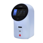
Frederick, USA: Syngene, a world-leading manufacturer of image analysis solutions, is delighted to announce its G:BOX imaging system is being used by scientists at a UK university to help visualize protein signals which will detect how cells in the human body react to new drugs.
Scientists at the university are using a high resolution G:BOX system to accurately analyze fluorescent and chemiluminescent proteins on Western blots. The system is also being used to visualize proteins on 1D and 2D protein gels stained with Coomassie blue and agarose gels of DNA stained with Sybr Safe™ and Ethidium bromide. The information from the gels and blots is being used to determine the effectiveness of new therapeutics, which could potentially speed up drug development. A researcher in the university commented: “We are using DNA-based reporter plasmids to help construct an integrated array of sensors. When a drug excites the sensors a unique
 protein expression signature pattern is produced and these are being used to compile a reference catalog of signature patterns. To study this protein expression, we run a large number of 1D and 2D protein gels so we need an easy to use, yet accurate imaging system.”t a UK university to help visualize protein signals which will detect how cells in the human body react to new drugs.The researcher added: “We reviewed systems from two other suppliers before we installed our G:BOX in 2008 but we were not able to achieve the level of 2D gel imaging and analysis we wanted with either system. In the time we have been using the G:BOX we have found we can easily view our blots and accurately quantify protein expression, without needing a manual and an hour to get up and running and this has helped us advance our project.”
Laura Sullivan, Syngene’s Divisional Manager, explained: “Thousands of drugs are tested each year but only a fraction are used to treat patients and this is why any new technology to reduce the time and expense of detecting those effective drugs is so important. We are delighted that our imaging system is playing a role in this project because it is proving that for high throughput analysis of 2D gels, a high resolution G:BOX system really does offer exceptional performance.”
Syngene systems are sold direct in New England exclusively through New England BioGroup, LLC.


