Agarose gel electrophoresis is a method used in biochemistry and molecular biology to separate a mixed population of DNA and RNA fragments by length, to estimate the size of DNA and RNA fragments or to separate proteins by charge.[1] Nucleic acid molecules are separated by applying an electric field to move the negatively charged molecules through an agarose matrix. Shorter molecules move faster and migrate farther than longer ones because shorter molecules migrate more easily through the pores of the gel. This phenomenon is called sieving.[2] Proteins are separated by charge in agarose because the pores of the gel are too large to sieve proteins.
Applications
- Estimation of the size of DNA molecules following restriction enzyme digestion, e.g. in restriction mapping of cloned DNA.
- Analysis of PCR products, e.g. in molecular genetic diagnosis or genetic fingerprinting
- Separation of restricted genomic DNA prior to Southern transfer, or of RNA prior to Northern transfer.
Agarose gels are easily cast and handled compared to other matrices and nucleic acids are not chemically altered during electrophoresis. Samples are also easily recovered. After the experiment is finished, the resulting gel can be stored in a plastic bag in a refrigerator.

How to make 1% agarose gel: In a small beaker, mix 32mL of purified H2O, 4mL of 10x TBE buffer or 10x whichever buffer going to be used in electrophoresis, and 0.4g of agarose powder. Heat on hot plate, or microwave, until solution is almost boiling (about 98 degrees Celsius). Immediately pour solution into electrophoresis mold or Galileo Bioscience gel casting tray with notch (well) comb intact, let settle for approximately 30 minutes, or until it appears firm. Be careful, a gel which is perfectly molded is still extremely fragile. Remove notch (well) comb carefully, but quickly. Wrap in plastic and place in refrigerator if gel is not for immediate use.
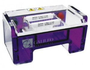
There are limits to electrophoretic techniques. Since passing current through a gel causes heating, gels may melt during electrophoresis. Electrophoresis is performed in buffer solutions to reduce pH changes due to the electric field, which is important because the charge of DNA and RNA depends on pH, but running for too long can exhaust the buffering capacity of the solution. Further, different preparations of genetic material may not migrate consistently with each other, for morphological or other reasons. Gel electrophoresis can be performed using a Galileo Bioscience gel electrophoresis system, or similar horizontal gel electrophoresis systems.
Factors affecting migration of nucleic acids
The most important factor is the length of the DNA molecule, smaller molecules travel faster. But conformation of the DNA molecule is also a factor. To avoid this problem linear molecules are usually separated, usually DNA fragments from a restriction digest, linear DNA PCR products, or RNAs.
Increasing the agarose concentration of a gel reduces the migration speed and enables separation of smaller DNA molecules. The higher the voltage, the faster the DNA moves. But voltage is limited by the fact that it heats the gel and ultimately causes it to melt. High voltages also decrease the resolution (above about 5 to 8 V/cm).
Conformations of a DNA plasmid that has not been cut with a restriction enzyme will move with different speeds (slowest to fastest: nicked or open circular, linearised, or supercoiled plasmid).
Visualization: ethidium bromide (EtBr) and dyes
The most common dye used to make DNA or RNA bands visible for agarose gel electrophoresis is ethidium bromide, usually abbreviated as EtBr. It fluoresces under UV light when intercalated into DNA (or RNA). By running DNA through an EtBr-treated gel and visualizing it with UV light, any band containing more than ~20 ng DNA becomes distinctly visible. EtBr is a known mutagen, however, so safer alternatives are available.
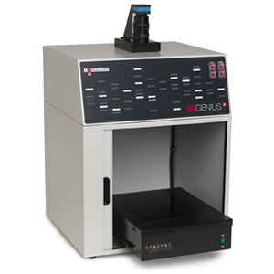
Even short exposure of nucleic acids to UV light causes significant damage to the sample. UV damage to the sample will reduce the efficiency of subsequent manipulation of the sample, such as ligation and cloning. If the DNA is to be used after separation on the agarose gel, it is best to avoid exposure to UV light by using a blue light excitation source such as the Ultraslim Blue Light Transilluminator or Ultra Bright Blue Light Transilluminator from Syngene and available in New England through New England BioGroup, LLC . A blue excitable stain is required, such as one of the SYBR Green or GelGreen stains. Gels with this stain can be visualized using a Syngene gel documentation system like the U:Genius, InGenius, GENi, and

G:BOX.
Blue light is also better for visualization since it is safer than UV (eye-protection is not such a critical requirement) and passes through transparent plastic and glass. This means that the staining will be brighter even if the excitation light goes through glass or plastic gel platforms.
SYBR Green I is another dsDNA stain, produced by Invitrogen. It is more expensive, but 25 times more sensitive, and possibly safer than EtBr, though there is no data addressing its mutagenicity or toxicity in humans.[3] Gels with this stain can be visualized using a Syngene gel documentation system like the U:Genius, InGenius, GENi, and G:BOX.
SYBR Safe is a variant of SYBR Green that has been shown to have low enough levels of mutagenicity and toxicity to be deemed nonhazardous waste under U.S. Federal regulations.[4] It has similar sensitivity levels to EtBr,[4] but, like SYBR Green, is significantly more expensive. In countries where safe disposal of hazardous waste is mandatory, the costs of EtBr disposal can easy outstrip the initial price difference, however.
Since EtBr stained DNA is not visible in natural light, scientists mix DNA with negatively charged loading buffers before adding the mixture to the gel. Loading buffers are useful because they are visible in natural light (as opposed to UV light for EtBr stained DNA), and they co-sediment with DNA (meaning they move at the same speed as DNA of a certain length). Xylene cyanol and Bromophenol blue are common loading buffers; they run about the same speed as DNA fragments that are 5000 bp and 300 bp in length respectively, but the precise position varies with percentage of the gel. Other less frequently used progress markers are Cresol Red and Orange G which run at about 125 bp and 50 bp.
Visualization can also be achieved by transferring DNA to a nitrocellulose membrane followed by exposure to a hybridization probe. This process is termed southern blotting.
Percent agarose and resolution limits
Agarose gel electrophoresis on a system like the Galileo Bioscience or Thermo Owl Scientific gel electrophoresis gel running system can be used for the separation of DNA fragments ranging from 50 base pair to several megabases (millions of bases) using specialized apparatus. The distance between DNA bands of a given length is determined by the percent agarose in the gel. The disadvantage of higher concentrations is the long run times (sometimes days). Instead high percentage agarose gels should be run with a pulsed field electrophoresis (PFE), or field inversion electrophoresis.
Most agarose gels are made with between 0.7% (good separation or resolution of large 5–10kb DNA fragments) and 2% (good resolution for small 0.2–1kb fragments) agarose dissolved in electrophoresis buffer. Up to 3% can be used for separating very tiny fragments but a vertical polyacrylamide gel is more appropriate in this case. Low percentage gels are very weak and may break when you try to lift them. High percentage gels are often brittle and do not set evenly. 1% gels are common for many applications.
Buffers
There are a number of buffers used for agarose electrophoresis. The most common being:Tris/Acetate/EDTA (TAE), Tris/Borate/EDTA (TBE) and lithium borate (LB). TAE has the lowest buffering capacity but provides the best resolution for larger DNA. This means a lower voltage and more time, but a better product. LB is relatively new and is ineffective in resolving fragments larger than 5 kbp; However, with its low conductivity, a much higher voltage could be used (up to 35 V/cm), which means a shorter analysis time for routine electrophoresis. As low as one base pair size difference could be resolved in 3 % agarose gel with an extremely low conductivity medium (1 mM Lithium borate).[5]
Analysis
After electrophoresis the gel is illuminated with an ultraviolet lamp (usually by placing it on a Syngene UV transilluminator light box, while using protective gear to limit exposure to ultraviolet radiation) to view the DNA bands. The ethidium bromide fluoresces reddish-orange in the presence of DNA. The DNA band can also be cut out of the gel, and can then be dissolved to retrieve the purified DNA. The gel can then be photographed usually with a digital or polaroid camera. Although the stained nucleic acid fluoresces reddish-orange, images are usually shown in black and white (see figures).
Gel electrophoresis research often takes advantage of software-based image analysis tools, such as Syngene GeneTools image analysis software, Nonlinear Dynamincs TotalLab or Phoretix software, NIH ImageJ, or Bio-Rad Image Lab or Quantity One image analysis software.
Typical method
Materials
Typically 10-30 μl/sample of the DNA fragments to separate are obtained, as well as a mixture of DNA fragments (usually 10-20) of known size (after processing with DNA size markers either from a commercial source or prepared manually).
- Buffer solution, usually TBE buffer or TAE 1.0x, pH 8.0
- Agarose
- An ultraviolet-fluorescent dye, ethidium bromide, (5.25 mg/ml in H2O). The stock solution be careful handling this. Alternative dyes may be used, such as SYBR Green.
- Nitrile rubber gloves. Latex gloves do not protect well from ethidium bromide
- A color marker dye containing a low molecular weight dye such as “bromophenol blue” (to enable tracking the progress of the electrophoresis) and glycerol (to make the DNA solution denser so it will sink into the wells of the gel).
- A gel rack such as a Bio-Rad, Owl Scientific or Galileo Bioscience horizontal gel electrophoresis running system.
- A “comb”
- Power Supply
- UV lamp or UV lightbox or a gel doc system, such as a Syngene, Bio-Rad, UVP, Carestream Healthcare or other gel documentation system to visualize DNA in the gel.
Preparation
There are several methods for preparing gels. A common example is shown here. Other methods might differ in the buffering system used, the sample size to be loaded, the total volume of the gel (typically thickness is kept to a constant amount while length and breadth are varied as needed). Most agarose gels used in modern biochemistry and molecular biology are prepared and run horizontally.
- Make a 1% agarose solution in 100ml TAE, for typical DNA fragments (see figures). A solution of up to 2-4% can be used if you analyze small DNA molecules, and for large molecules, a solution as low as 0.7% can be used.
- Carefully bring the solution just to the boil to dissolve the agarose, preferably in amicrowave oven.
- Let the solution cool down to about 60 °C at room temperature, or water bath. Stir or swirl the solution while cooling.
Wear gloves from here on, ethidium bromide is a mutagen!
- Add 5 µl ethidium bromide (from a stock solution of 10 mg/ml) per 100 ml gel solution for a final concentration of 0.5 µg/ml. Be very careful when handling the concentrated stock. Some researchers prefer not to add ethidium bromide to the gel itself, instead soaking the gel in an ethidium bromide solution after running, in this case the ethidium bromide solution has final concentration of 0.5 µg/ml.
- Stir the solution to disperse the ethidium bromide, then pour it into the gel rack.
- Insert the comb at one side of the gel, about 5–10 mm from the end of the gel.
- When the gel has cooled down and become solid, carefully remove the comb. The holes that remain in the gel are the wells or slots.
- Put the gel, together with the rack, into a tank with TAE. Ethidium bromide at the same concentration can be added to the buffer. The gel must be completely covered with TAE, with the slots at the end electrode that will have the negative current.
Procedure
After the gel has been prepared, use a micropipette to inject about 2.5 µl of stained DNA (a DNA ladder is also highly recommended). Close the lid of the electrophoresis chamber and apply current (typically 100 V for 30 minutes with 15 ml of gel). The colored dye in the DNA ladder and DNA samples acts as a “front wave” that runs faster than the DNA itself. When the “front wave” approaches the end of the gel, the current is stopped. The DNA is stained with ethidium bromide, and is then visible under ultraviolet light.
- The agarose gel with three slots/wells (S).
- Injection of DNA ladder (molecular weight markers) into the first slot.
- DNA ladder injected. Injection of samples into the second and third slot.
- A current is applied. The DNA moves toward the positive anode due to the negative charges on its phosphate backbone.
- Small DNA strands move fast, large DNA strands move slowly through the gel. The DNA is not normally visible during this process, so the marker dye is added to the DNA to avoid the DNA being run entirely off the gel. The marker dye has a low molecular weight, and migrates faster than the DNA, so as long as the marker has not run past the end of the gel, the DNA will still be in the gel.
- Add the color marker dye to the DNA ladder.
References
Kryndushkin DS, Alexandrov IM, Ter-Avanesyan MD, Kushnirov VV (2003). “Yeast [PSI+] prion aggregates are formed by small Sup35 polymers fragmented by Hsp104”. Journal of Biological Chemistry 278: 49636. doi:10.1074/jbc.M307996200.
Sambrook J, Russel DW (2001). Molecular Cloning: A Laboratory Manual 3rd Ed. Cold Spring Harbor Laboratory Press. Cold Spring Harbor, NY.
SYBR Green I Nucleic Acid Gel Stain
4.0 4.1 SYBR Safe DNA Gel Stain
↑ Brody, J.R., Kern, S.E. (2004): History and principles of conductive media for standard DNA electrophoresis. Anal Biochem. 333(1):1-13. PMID 15351274 PDF
Copyright Information
This article is distributed under the Creative Commons Attribution/Share-Alike License. For information on the contributors, please see the original Wikipedia article. It has been adapted and modified for commercial use by New England BioGroup, LLC under the Creative Commons Legal Code (Full License).


