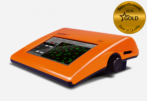
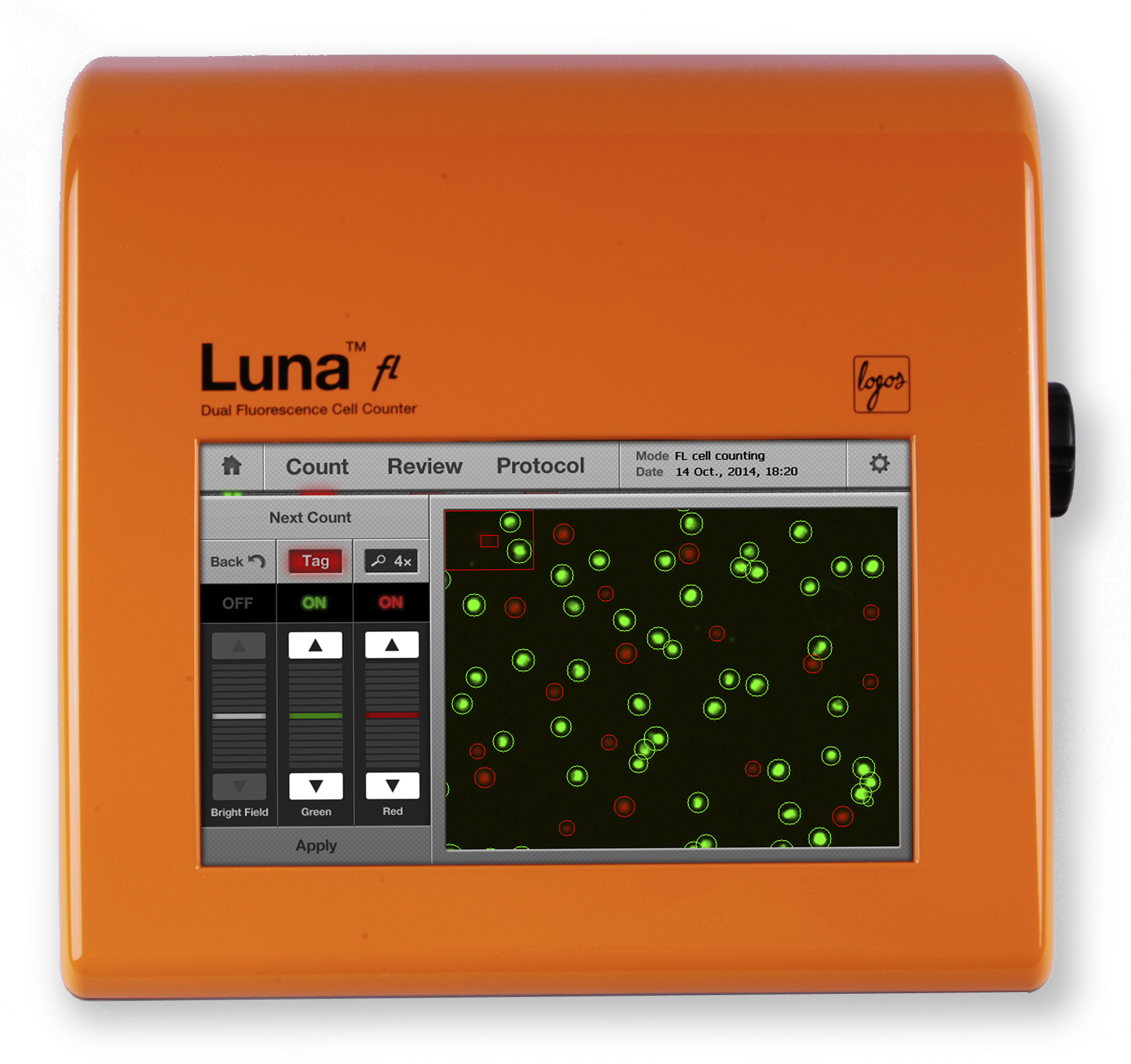
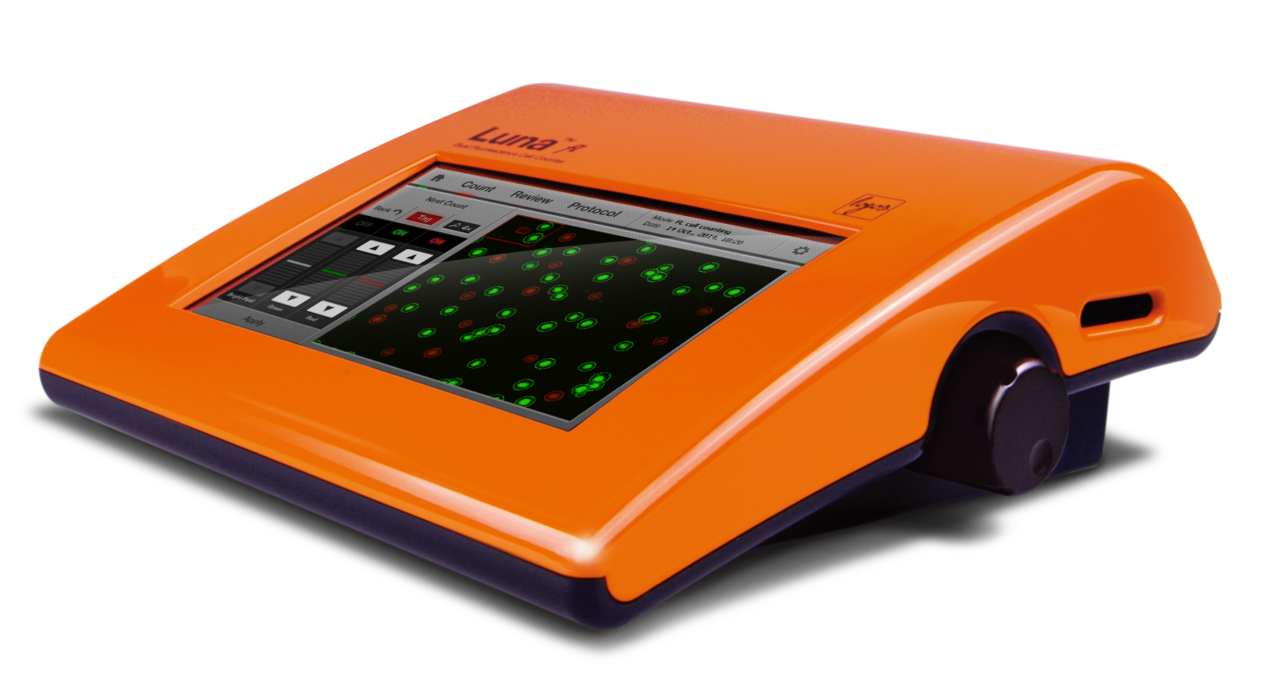
The LUNA-FL™ Dual Fluorescence Cell Counter from Logos Biosystems is a quantum leap forward for automated cell counting and cell viability analysis. The LUNA-FL™ Dual Fluorescence Cell Counter gives you sensitive and accurate live/dead counts without cell type restrictions. Unlike immortalized cell lines, primary cells such as PBMCs, splenocytes, neutrophils, and stem cells have been difficult to count with conventional cell counters such as the Coulter counter or automated image-based cell counters utilizing bright field optics. Primary cells are often contaminated with undesirable debris that can be miscounted as cells with conventional cell counters. The LUNA-FL™ is equipped with dual fluorescence optics to overcome this problem. Live and dead cells are stained with green and red fluorescence dye, respectively, and subsequent cell images are analyzed with accurate image analyzing software
To read a general review of fluorescent cell counting, please click here.

The LUNA-FL™ has inherited the proven performance of the LUNA™. The precision bright field optics of the LUNA™ has been integrated into the LUNA-FL™ to provide the convenience of trypan blue cell counting. The powerful and precise cell counting algorithm of the LUNA™ is used in the LUNA-FL™ LUNA-FL™ = LUNA™ + Dual fluorescence cell counter
The LUNA-II™ automated cell counting algorithm has been recognized to have the best-in-class cell counting accuracy. The LUNA™ software has exceptional acc
uracy in cell de-clustering and can successfully count clumpy cells. The LUNA-II™ automated cell counter has inherited this well-known performance aspect of the LUNA™ software. Clumpy cells are de-clustered quickly and automatically, then counted as individual cells.

With the LUNA-FL™ Dual Fluorescence Cell Counter, GFP expression analysis takes less than 30 seconds. Bright field and fluorescence cell images are analyzed to detect GFP-positive and negative cells, and a corresponding fluorescence intensity histogram is displayed for further analysis. The LUNA-FL™ Dual Fluorescence Cell Counter is integrated with on-board flow cytometry type gating software. Be confident in the accuracy of your results by adjusting the threshold of fluorescence intensity.
Automated cell counters utilize disposable counting slides to eliminate washing steps of manual cell counting with the glass hemocytometer. Although disposable cell counting slides have several advantages, the increased running cost has been a substantial concern. Logos Biosystems developed a patented T-BOND technology to manufacture precision cell counting slides more efficiently. Therefore, the unit price of counting slides became much more affordable, providing significant cost savings.
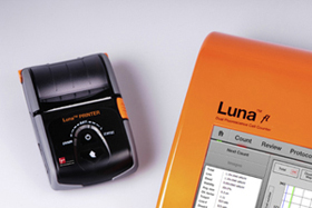 Compact and Fast Thermal Printer (optional)
LUNA-FL™ provides a compact mobile printer for further record keeping purpose. Thermal roll paper or label paper can be selected. This plug & play mobile printer is ready-to-use after the USB to USB connection.
The LUNA-II™ automated cell counter can count samples with or without trypan blue dye. The selection of trypan blue presence
or absence can easily be chosen in the settings menu.
After counting, cells can be gated based on their size information provided by the LUNA-II™. The specific size of cell populations
Can easily be included or excluded on histograms.
Counted cells can be sorted with cell cluster map, into single cells, doublets, or triplets. The cell cluster map can be used to monitor changes in culture conditions or cell isolation/preparation protocols.
The LUNA-II™ automated cell counter provides a powerful review option. A separate PC unnecessary. Users can open cell images directly on the LUNA-II™ automated cell counter to check previous results.
The LUNA-II™ can store up to 1,000 counts onboard. After counting, the result is automatically saved in the memory, and previous count results can be exported via USB as a .CSV file for further analysis. The results and image data can be saved in three different file formats such as TIF, annotated TIF, or a PDF report file. The PDF report file contains all the data generated during/after the cell counting, i.e., the protocol used, cell counting results, date, raw/analyzed cell image(s), and histograms are included in the PDF report file.